Sunday, 23 February 2020

Unit 5 Sex determination


Unit 5
UNIT-  5


SEX DETERMINATION
S y l l a b u s
Chromosomal mechanisms and methods
(02 Lectures)

Chromosomal mechanisms and methods
Members of almost all species are often divided into two sections according to the kinds of gametes of sex cells produced by them i.e. male sex and female sex. The word sex has been derived from Latin word sexus meaning section or separation. However, some of the lowest forms of plant and animals like are found to have several sexes. For example, in one variety of ciliated protozoan, Paramecium bursaria there are eight sexes or ‘mating types’all morphologically identical. Each mating type is physiologically capable of conjugating with its own type, but may exchange genetic material with any of the seven other types within the same variety. Further, in organisms in which the number of sex reduced to just two, sexes may reside in different individuals or within the same individual. An animal possessing both male and female reproductive organs is usually referred to as a hermaphrodite. The sex cells and reproductive organs form the primary sexual characters of male and female sexes. Besides these primary sexual characters, the male and female sexes differ from each other in many somatic characters known as secondary sexual characters. The phenomenon of molecular, morphological, physiological or behavioral differentiation between male and females sexes is called sexual dimorphism.
The phenomenon of sexual dimorphism has been a biological riddle for the thinkers and biologists of all time. People always tried to know those factors which determine the male and female sexes of a species. Literally hundreds of mistaken hypotheses and wild guesses were proposed before 1900 in vain attempts to find out a solution to the problem of determination of sex. Modern genetics have reported many different mechanisms of determination of sex in living organisms.
The X chromosomes have large amount of euchromatin and small amount of heterochromatin as compared to Y chromosome. The euchromatin has large amount of DNA material, hence, X chromosomes is genetically active.  The Y chromosome contains small amount of euchromatin and large amount of heterochromatin. The chromosome has little genetic information; therefore, sometimes it is referred to as genetically inert or inactive chromosome.
Sex Determination in Animals:
Biologically, sex is an aggregate of those morphological, Physiological and behavioral qualities that differentiates the organisms producing eggs from those producing sperms. This separation of species or section of animals into two sexes is called as Gonochorism. The sex chromosome behaves as a Mendelian inheritance follows the law of segregation. Many mechanisms are given to explain the determination and differentiation of sexes.
These mechanisms are –
A) Genetically controlled sex determining mechanism
i)        Chromosomal theory of sex determination or sex chromosome mechanism
ii)      Genic balance mechanism
iii)    Haplodiploidy mechanism
iv)    Single gene effects or single gene control of sex
Among these sex chromosomes, theory of sex determination is best explanatory in explaining the problem of sex determination. In plants male and female do not differ morphologically, except in the floral characters and here gonochorism is a rare phenomenon.
i) Chromosomal Theory of Sex determination or Sex Chromosomal Mechanisms or Heterogamesis:
a)   According to the chromosome theory of sex determination the male (♂) and female (♀) individuals normally differ in their chromosomal constituents. There may be two types of chromosomes present in these individuals, theseare :
            Sex chromosomes or Allosomes,i.e., chromosomes, which are responsible for determination of sex, e.g., X and Y-chromosomes.
            Autosomes,i.e., chromosomes, which have no relation with the sex and contain genes, which determine the somatic characters.
b)   The X-chromosome was first observed by German biologist Henking in 1891, during his studies on spermatogenesis in male bug. He called it X-body.
c)   The chromosomal theory of sex determination was worked out by E. B. Wilson and Stevens (1902-1905). They named X and Y chromosomes as allosomes or sex chromosomes and other chromosomes as autosomes.
d)   The parents can be of two types -
      •     Homogametic, i.e., with similar gametes.
      •     Heterogametic, i.e., with different gametes.
e)   In some cases, males are heterogametic, e.g., Man, Drosophila, bug, etc., while in some other cases, females are heterogametic, e.g., birds. Thus, we have two following types of systems among diploid organisms, on the basis of chromosomal theory of sex determination.
i.        System that is having heterogametic males.
ii.      System that is having heterogametic females.
f)   In the first system, i.e., males heterogametic, the females have two X-chromosomes, while males have one X- chromosome and Y- chromosome because of which male during gametogenesis produces two types of gametes (sperms), i.e.,
•     50% that carry X- chromosome.
•     50% that carry Y- chromosome.
g)   This chromosomal pattern varies in different organisms in the way that male either contain Y-chromosome (i.e., XX - XY system or lygaeus type) or contain no chromosome along with X- chromosome, i.e., XX - XO type or protenor type.
Structure of sex chromosomes
The X and Y chromosomes exhibit structural differences. The cytological studies have shown that the X chromosomes of most organisms are straight, rod-like and comparatively larger than Y chromosomes. The Y chromosome is smaller in size, with one end slightly curved or bent to one side in Drosophila; in man and Melandrium no such curvature of Y chromosome occurs. The X chromosome has large amount of euchromatin and small amount of heterochromatin. The euchromatin has large amount of DNA material, much genetic information. The Y chromosome contains small amount of euchromatin and large amount of heterochromatin. The Y chromosome has little amount of genetic information, therefore, sometimes it is referred to as genetically inert or inactive.


Fig. Male and Female Drosophila melanogaster and their chromosome
Discovery of sex chromosomes
In sexually dimorphic dioecious organisms, besides morphological and behavioral differences between both sexes, the sexual diversity also occurs at the level of chromosomes. The chromosomal differences between the sexes of several dioecious species were found earlier in the course of cytological investigations. A German biologist, Herman Henking in 1891 while studying spermatogenesis of the squash bug, Pyrrhocoris, noted that meiotic nuclei contained 11 pairs of chromosomes and an unpaired element is moved to one of the poles during the first meiotic division. Henking called this unpaired element a ‘X-body’ and interpreted it as a nucleolus. The significance of X-body was not immediately understood, but in 1902 an American geneticist, McClung, who had made extensive observations of gametogenesis in grasshoppers, suggested that the X-body was involved in some way with the determination of sex. He reported that somatic cells of the female grasshopper (Xyphidium faciatum) contained 24 chromosomes, whereas those of the male had only 23. He demonstrated that karyotype of a cell is composed not only of common chromosomes (autosomes) but also of one or more special chromosomes that are distinguished from the autosomes by their morphological characteristics and behavior. These were called accessory chromosomes, allosomes, heterochromosomes or sex chromosomes. In 1905, Edmond Wilson noted that females of Protenor, a hemipteran bug have 7 pairs of chromosomes, while the males have 6 pairs and an unpaired chromosome, which he called the X chromosome. The X and Y chromosomes were first discovered in beetles by Nettie Stevens in 1906. Stevens found a similar situation in Drosophila melanogaster, which has four pairs of chromosomes, with one of the pairs being heteromorphic in nature.


Fig.  Sex determination pathways in diverse model organisms.
Types of Sex Chromosomal Mechanism of Sex Determination:
In dioecious diploidic organisms following two systems of sex chromosomal determination of sex have been recognized;
(a) Heterogametic males and (b) Heterogametic females
a) Heterogametic Males:
In this type of sex chromosomal determination of sex, the female sex has two X-chromosomes, while the male sex has only one X chromosome. Because, male lacks a X chromosome, therefore, during gametogenesis it produces two types of gametes, 50 per cent gametes carry the X-chromosomes, while the rest lack in X chromosomes.
Such as sex which produces two different types of gametes in terms of sex chromosomes is called heterogametic sex. The female sex is therefore called homogametic sex. The heterogametic males may be of the following two types.
i) XX - XY method: The XX - XY method is the most common method of sex determination. It is found in mammals and certain insects like Drosophila, etc. In these animals the female possesses two homomorphic X- chromosomes (i.e., XX) and being homogametic produces eggs of one kind only. These all eggs have one X- chromosome in them.
The male, here possesses one X and one Y- chromosome (hence, called XY) and produces two kinds of sperms, i.e., 50% with X- chromosome and 50% with Y- chromosome.
The sex      of embryo depends upon the kind of sperm. A female individual is produced, if egg fertilizes with a sperm having X-chromosome and a male is produced if same is fertilized with Y-chromosome bearing sperm.
XX-XY method (Example man and Drosophila)
In human being there are 46 chromosomes (23pairs) in all the body cells except gametes. Of these 46 chromosomes, 44 are autosomes which are common in both the sexes and the remaining two are sex chromosomes either with XX combination or with XY combination. The female has 44 autosomes and two sex chromosomes- XX; the male has 44 autosomes and two sex chromosomes- XY. Thus combination of sex chromosomes determines the sex, two XX chromosomes gives rise to a female whereas X and Y combination gives rise to a male.


                                                                                     Fig. XX - XY pattern
ii) XX – XO pattern - The other method, i.e., XX – XO type is found in certain insects especially those of class – Hemiptera (true bugs) and order – Orthoptera, In this method, the female has 2X–chromosomes (called while male has only one X– chromosome (called XO).
The Y–chromosome is completely lacking here, thus, the presence of unpaired X – chromosome determines masculine sex. The female just like lygaeus method produces only one type of eggs and male produces types of sperms, i.e., 50% with one X – chromosome and 50% without any sex chromosome. The sex of offspring depends upon the type of sperm, which fertilizes the egg, e.g., in grasshopper.
Example Grasshopper In 1902 C. E. McClung while studying meiosis in the testes of the Grasshopper, he noted that there were 11 pairs (22) of chromosomes and an odd chromosome with no mate. He associated the odd chromosome with the sex determination. Thus the female grasshopper possesses 22 autosomes and a pair of sex chromosomes i.e. XX and the male possesses 22 autosomes and only one sex chromosome, the X.  The female is homogametic and produces only one kind of egg with 11 autosomes and one sex chromosome, the X. The male is heterogametic and produces two types of sperms one with 11 autosomes and one sex chromosome, X and other with 11 autosomes only (that is without a sex chromosome). The sex determination in grasshopper follows a different pattern as illustrated below;
b) Heterogametic Females
In this type of sex chromosomal determination of sex, the male sex possesses two homomorphic X chromosomes, therefore, is homogametic and produces single type of gametes, each carries a single X chromosome. The female sex either consists of single X chromosome or one X chromosome and one Y chromosome. The female sex is, thus, heterogametic and produces two types of eggs, half with a X chromosome and half without a X chromosome (with or without a Y chromosome). To avoid confusion with that of XX-XO and XX-XY types of sex determining mechanisms, instead of the X and Y alphabets, Z and W alphabets are generally used respectively. The heterogametic females may be of following two types:


Fig. Haploid male, Diploid female and XX-XO type
sex determination mechanism of insects.
i)        ZO-ZZ method (Example butterflies and moth)
This system of sex determination is found in certain moths and butterflies. In this case, the female possesses single Z chromosome in its body cells (hence, is referred to as ZO) and is heterogametic, producing two kinds of eggs, half with a Z chromosome and half without any Z chromosome. The male possesses two Z chromosomes (hence, referred to as ZZ) and is homogametic, producing single type of sperms, each of which carries a single Z chromosome. The sex of the offspring depends on the kind of egg as shown below;


Fig. ZO-ZZ determination of sex in butterfly
ii) ZZ-ZW method (Example Birds)
A number of animals display a method of sex determination wherein the relation of the sex chromosomes is exactly the reverse of the XY or the XO method. The rooster (male fowl) would have two like sex chromosomes designated as ZZ and would produce only one type of sperms, while a hen would have two unlike sex chromosomes ZW and would produce two types of eggs. Sperms would all carry a Z chromosome but the eggs would carry either Z or W chromosomes in equal numbers, sex determination therefore depends upon which of the two types of eggs is fertilized.


Fig The ZW-ZZ type of determination of sex in chicken
ii) Genic Balance Mechanism:
  • The theory of genic balance given by Calvin Bridges (1926) states that instead of XY chromo­somes, sex is determined by the genic balance or ratio between X-chromosomes and autosome genomes.
  • The sex determination in Drosophila is quite different from humans. Drosophila has eight chromosomes (n = 4), three pairs of autosomes and one pair of sex chromosomes. Even though Drosophila possesses XX and XY sex chromosomal organization, unlike human beings, the Y chromosome does not have any role in determining the sex of individuals. The sex in Drosophila is determined by the ratio of the number of X chromosomes to that of the number of sets of autosomes. In simpler terms, the sex determination is achieved by a balance of female determinants on the X chromosome (X) and male determinants on the autosomes (A). This type of sex determination is called genic balance system.
Chromosome Complement
X / A Ratio
Sexual Morphology
X X X + 2A
3/2 or 1.5
Metafemale
X X X + ЗА
3/3 or 1.0
Female
XX + 2A
2/2 or 1.0
Female
X X + ЗА
2/3 or 0.67
Inter sex
X X X + 4A
3/4 or 0.75
Inter sex
XO + 2A
1/2 or 0.5
Male
XY + 2A
1/2 or 0.5
Male
XY + ЗА
1/3 or 0.33
Metamale
a) Triploid or polyploid intersexes in Drosophila
In many cases of different organisms, it has been observed that these generally have inherent potentialities both the sexes and each individual is found to be more or less intermediate between male and female sex. In Drosophila, in fact, it is the ratio between the X–chromosome and autosome, which governs or determines sex. This was worked by CB Bridges (1922), on triploid Drosophila.
He performed experiments on these triploids, on the basis of which he developed his ratio theory of sex determination or ultimately the genic balance theory. According to Bridges in Drosophila, it is the
a)   X–chromosome – Which is responsible for female characteristics.    
b)   Y–chromosome – Which is responsible for male characteristics.
c)   Autosome – It is necessary for the fertility of sex, but not responsible for any sex determination.
Thus, according to him, it is the delicate balance between the X–chromosomes and autosomes, which is responsible for sex determination. This theory is very helpful in predicting the sex of individuals that arise from non-disjunction of X-chromosome during meiosis in female. According to this theory, a singleX–chromosome in a diploid organism produces male sex and XX combination within a diploid organism produces females. In his experiments, Bridges crossed a triploid female (3A + XXX) with a diploid male (2A + XY), the result of this cross can be seen as


Fig. Classical cross of a triploid (3A+XXX) female fly and a diploid (2A+XY) male fly (Drosophila)
The presence of triploid inter sexes in the above experiment is a proof that autosomes also carry genes for sex determination. The intersexes, super males and super females were obtained,where interpreted by bridges in the form genic balance theory of sex determination. According to this theory, ratio between the number of X–chromosomes and number of complete sets of autosomes will determine sex.
If the ratio between X and A is 1.0, it will be female. If 0.5 it will be male, ifmore then 1, it will be super female, if less then 0.5, it will be super male and Ifbetween 0.5 to 1, it will be inter sex.
Table  Different doses of X-chromosomes and autosome sets and their effect on sex determination in Drosophila.
Phenotypes
Number of chromosomes (X)
Number of autosomes
(A sets)
Sex index =
Super female
3
2
1.5
Normal female
1)      Tetraploid
2)      Triploid
3)      Diploid
4)      Haploid
4
3
2
1
4
3
2
1
1.0
1.0
1.0
1.0
Intersex
2
3
0.67
Normal male
1
2
0.50
Super male
1
3
0.33

b)  Gynandromorphs in Drosophila.
In Drosophila, occasionally, flies are obtained with one half having male characteristics and another half having female characteristics
Concept of sex determination as developed for Drosophila is verified by the occasional occurrence of gynandromorphs which are individual in which part of the body expressed male characters, whereas other parts express female characters. In a way, gynandromorphs represent one kind of mosaic or an organism made up of tissues of male and female genotypes. For example, bilateral gynandromorphs of Drosophila are male on one side (either right or left) and female on the other. It results due to the loss of an X-chromosome in a particular cell during development i.e. when the pair of sex X-chromosome fails to separate during gametogenesis one X and is lost forever. If this event happens during first cleavage of mitotic division or the zygote, then one of the two blastomeres will have AAXX chromosomal complement and the other will have AAXO. The portion of the body developing from AAXX blastomeres will be normal female and the portion developing from the AAXO blastomeres will be male. The cytological examination of gynandromorphs suggested that Y-chromosome does not play any role in the determination of sex in Drosophila. , as shown below


Fig.7.5:Gynandromorph of Drosophila, in which right half is male and left half is female


Fig. The loss of an X chromosome during mitosis in a 2A+XX cell and formation of two types of cells – XX and XO

iii) Haplodiploidy Mechanism:
Another mechanism of sex determination is male haploidy or haplodiploidy. This mechanism is particularly common in hymenopteran insects, e.g., honey bee, wasp, ants, etc.
The pattern is more common in those insects, in which parthenogenesis is generally seen. In these insects, normally, three types of individuals are seen -
a) Diploid queen: Fully functional females developed from fertilized eggs.
b) Diploid workers:Underdeveloped or non-functional females developed from fertilized eggs.
Actually here, the fertilized egg gives the females in general. Out of which, the female which fed on Royal jelly (a specialized food) becomes queen and rest are known as workers.
The workers have specific function of protection of colony, while queen involved in egg production mainly. The workers are unable to perform fertilization and egg laying as in them, the organs of ovipositionare mo6dified to sting apparatus.
c) Drones: Functional males are developed parthenogenetically from unfertilized haploid eggs. Drones are haploid organism. These males are short lived and normally die after fertilization.
As males are developed parthenogenetically, hence, we can say that in these mechanisms, parthenogenesis (i.e., development of eggs to adult without fertilization) plays a major role in determination of the sex. The sex chromosomes have no identity here.  In certain extreme cases, males are altogether absent and females are produced by parthenogenesis. These females are always diploid. e.g., Lacertasexicota. In Habrobraconjuglandis (a parasitic wasp also called Braconhebetor) the sex determination mechanism is similar to wasps and bees, i.e., unfertilized eggs develop into males and fertilized eggs develop into females.
The difference lies in the fact that some fertilized eggs also develop into males. This is due to the presence of a number of alleles promoting maleness and femaleness, e.g., XA, XB, XC and XD, etc. The diploid males have homozygous condition of these alleles, while diploid females have heterozygous condition. 
Honeybee or Diploid-haploid method of sex determination
The honeybee and certain other hymenopterans possess a peculiar type of sex determination. These insects can produce either sex at will. In other words honeybees and other such insects can determine the sex of their progeny at their own will. This is a unique example wherein mother can decide the sex of her child. The queen leaves the hive on her nuptial flight and flies high in the air, pursued by the drones from her own and surrounding hives. Copulation occurs during her nuptial flight and sperms are transferred by a drone to a queen’s body. These sperms are stored in a seminal vesicle within her body and remain available for fertilization when eggs are laid throughout remainder of her life. Now the queen can control the fertilization of egg and can lay either fertilized eggs, by either allowing sperms to come in contact with the eggs or by preventing sperm contact with eggs respectively. The fertilized eggs develop into males. Thus honeybee method of sex determination is quite distinct from the other chromosomal methods.


 Fig. Diploid-Haploid Sex determination system


iv) Single gene effects or single gene control of sex
In certain organisms, for example Chlamydomonas, Neurospora, yeast, Asparagus, maize, Drosophila, etc., individual single genes are found to be responsible for the determination or expression of sex, following cases exemplified the single gene control of sex:
a)   Monogenic sex determination in Drosophila: In Drosophila, a transformer gene (tra) has been recognized which when present in homozygous condition (tra/ tra) transforms a female fly into a sterile male, but, it does not act upon normal male individuals. Thus, a XX female with tra/ tra genotype will be a sterile male, but, a XY male with tra/ tra genotype will still be a normal male fly.
b)   Sex reversal gene (Sxr) in mammals: Recently a sex reversal gene (Sxr) has been discovered in human beings, so that in the presence of this gene XX female individuals may become male. Such cases of sex reversal are also reported in goat and mice. Mice also contain two other genes Tdyand Tda-1 which interact to cause sex reversal in XY male individuals to transform them into females.
c)   Complementary sex factors: Besides the haploidy mechanism of sex determination, two hymenopterans insects – Bracon hebetor (parasitic wasp) and honeybees are known to produce males by homozygocity at a single gene locus.
d)   Sex-determination in Asparagus:Asparagus is a dioecious plant, however, sometimes the female flowers bear rudimentary anthers and the male flowers bear rudimentary pistils. Thus, sometime it may happen that a rare male flower with poorly develop pistil may set seeds. In one of the experiments when the seeds of such a rare male flower were raised into plants, then, the male and female plants were found to be present in 3: 1 ratio. When the male plants raised thus were used to pollinate the female flowers on female plants, only two third of them showed segregation indicating that the sex is controlled by a single gene.
B) Metabolically controlled sex determining mechanism:
Certain workers have seen the possibility of sex determination in the phenomenon of metabolism. Cruw suggested that sex is a physiological equitable division between anabolic and catabolic individuals. A.F. Shull and D.D. Whitney have shown that by increasing metabolic rate in rotifers the occurrence of male individuals increase than females.
C) Hormonal and Environmental Control of Sex Determination:
Besides above written mechanisms of genetic control, hormonal and environmental role is also seen in determining the sex. The sex determination theories (chromosomal theory and genic balance) apply to lower animals but in higher vertebrates, the hormones especially the gonadal hormones (secondary products of sex chromosomes) may considerably alter the sexual characters.
In a particular sex, a particular hormone produced by endocrine glands (gonads for normal development. The intentional or unintentional removal of such a gland results into changes in characteristic.
For example, in fowl, if the ovary from a genetic female is removed (intentionally or accidentally it results into the development of male comb along with male plumage and in extreme cases testes will also appear (findings of crene).
The above case of sex reversal was explained by assuming that as soon as the ovary was removed or destroyed, the ovarian hormones were stopped. After sometime, the dormant testes present as rudiments in almost all the female birds start functioning.
The male hormones were produced, which helped in the development of secondary sexual characters and sperm formation in extreme cases. Cases of such sex reversal are observed in some fishes, amphibians, birds and even in mammals.
In Bonelia viridis, all the larvae are genetically and cytologically similar. If a particular larva settles near proboscis of an adult female, becomes a male individual.
If it develops free in water, it becomes a female this is called environmental control of sex determination.

Unit 4 Mutation

Unit 4 Mutation

Chromosomal Mutation: Deletion, Duplication, Inversion, Translocations, Aneuploidy and polyploidy, Gene mutations Induced versus spontaneous mutations.
The Chromosomes of every species possess a definite number of chromosomes. Each chromosome has a specific structure. Which imparts, a specific phenotype. But during the cell division due to certain accidents or irregularities the alterations in chromosome number and its morphology takes place which changes the phenotype. It changes the original structure and number of chromosomes. Such type of change in genome involving chromosome parts or whole chromosome or a set of chromosome is called as chromosomal mutations or chromosomal aberrations.
Chromosomal mutations:
I) Structural changes in chromosomes
  • Changes in number of genes i.e. a) loss or deletion and b) addition or duplication
a) Deletion: Loss – Changes in number of genes. Loss of portion of chromosome is called deletion. Portions of the chromosomes without a centromere lag in anaphase movement and are lost from reorganizing nuclei or digested by nucleases. The chromosomes with deletions can never come back to normal condition. If gametes arise from the cells having a deleted chromosome this deletion is transmitted to the next generation.
The deletion is the breakage of chromosome at any position, it can be terminal or intercalary deletion.
Intercalary deletion:
Breakage takes place at the middle of chromosome. It involves two breakages in the chromosome.
Terminal Deletion:When breakage takes place at the end of chromosome, there is only single breakage in chromosome.
Example of deletions:
1)         Human babies missing a portion of the short arm of chromosome 5 (outosome) have a distinctive cat like cry called as “cri-du-chat” (cry of the cat) syndrome
2)         Turner syndrome: deletion of part of the short arm of one X- chromosome.
b) Duplications:
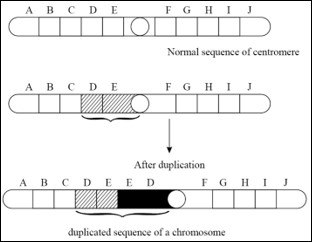
The presence of a part of a chromosome in excess of the normal complement is known as the duplication. Due to duplication some genes are present in more than two doses. If duplication is present only on one of the two homologous chromosomes during the meiosis the chromosome bearing the duplicated segment forms of loop to maximize the pairing of homologous regions.


 Types of duplications:
Tandem duplication: In this type the duplicated region is situated just by the side of the normal corresponding section of the chromosome and the sequences of genes are the same in normal and duplicated region
2. Reverse tandem duplication: In this type of duplication, the sequence of genes in the duplicated region of a chromosome is just the reverse of normal sequence.
 3) Displaced duplication:
In this type the duplicated region is not situated adjacent to the normal section. In this case the duplicated sequence is on the same side of the centromere as the original section or on the other side it may termed as homobranchial or heterobranchial


 Displaced duplication heterobranchial on different arm.
4) Transposed duplication: In this type of duplication, the duplicated portion of chromosome becomes attached to different i.e. non-homologous chromosome.
Examples of duplication:
Pallister Killian syndrome is a result of extra #12 chromosome material. There is usually a mixture of cells (mosaicism), some with extra #12 material, and some that are normal (46 chromosomes without the extra #12 material). Babies with this syndrome have many problems. These include severe intellectual disability, poor muscle tone, "coarse" facial features, and a prominent forehead. They tend to have a very thin upper lip, with a thicker lower lip and a short nose. Other health problems include seizures, poor feeding, stiff joints, cataracts in adulthood, hearing loss, and heart defects. People with Pallister Killian have a shortened lifespan, but may live into their 40s.


 Fig. Transposed duplication

1)      Bar eye in drosophila: This phenotype is characterized by narrower, oblong, bar shaped eye with few facets. It is due to duplication of a segment of the X chromosome, called section 16A.
2)      In humans. Concerning the hemoglobin gene, deletions result in lepore and Kenya variants of adult hemoglobin (Hba) both causing anemia.

  • Changes in gene arrangement:
When gene arrangement takes place Genes rotate within one chromosome or exchange of parts of chromosomes takes place between chromosomes.
1) Inversion:
It involves a rotation of a part of a chromosome or a set of genes by 1800 on its own axis. Here breakage and reunion of chromosomes takes place. Because of this there is neither a gain nor a loss in genetic material but it is simply a rearrangement of the gene sequence.
Inversion is of following two types.
The location of the centromere relative to inverted segment determines the genetic behavior of the chromosomes.
It is of two types.

1. Para centric inversion: When centromere is not included in the inversion, it is called paracentric inversion.

2)         Pericentric inversion: When inversion involves centromere it is called pericentric inversion
Advantages of inversions : Fertility of inversion homozygotes and sterility of inversion heterozygotes leads to establishment of two group (or varieties) which are mutually fertile but do not breed well with the rest of the species.
2) Translocations:
The transfer of a part of chromosome or a set of genes to a non-homologous chromosome is called as translocation.
The rearrangement of genes takes place The sequence and position of gene changes. There is no addition or loss of gene during translocations.
It is of following types :
a) Simple translocations:
It involves a single break in chromosome. The broken piece gets attached to one end of a non-homologous chromosome.
  b) Shift translocation:
In this type of translocation, the broken segment of one chromosome get inserted interstitially in a non-homologous chromosome.
c) Reciprocal translocation:In this  type, a segment from one chromosome is get exchanged with a segment from another non-homologous chromosome, In this way two translocation chromosomes are simultaneously achieved.
Example:
Philadelphia chromosome:
A translocation between the long arms of chromosome g and 22 often in the white blood cells of patients with chronic myeloid leukemia
II) Changes in number of chromosomes: It includes;
1.      Loss or gain of the part of the chromosome set (aneuploidy)
2.      Loss or gain of whole chromosome set (euploidy)
The phenomenon of variations in the number of chromosomes is called heteroploidy. It is of two types, euploidy and aneuploidy.
A) Euploidy
In this condition, an organism either loses a complete set of chromosomes or acquires one or more additional sets of chromosomes over and above the two sets of diploid complement. On this basis, euploidy can be categorized into two types – monoploidy (haploidy) and polyploidy.
a) Monoploidy or haploidy
Monoploidy have only one set of chromosomes. Haploids have half the somatic chromosome number. In diploid organisms, monoploids and haploids are identical while they differ in tetra or hexaploid organisms.
In certain organisms (e.g. honeybee) haploid offspring are produced as a routine which arise parthenogenetically from the unfertilized eggs. Haploids can be artificially induced by many methods like X-ray treatment, delayed pollination, culturing pollen grain and temperature shocks.
Haploids are smaller in size as compared to diplods. Their leaves, flowers, fruit and seeds are comparatively small. They are also less resistant and comparatively weak. The haploids have just univalent without any homologous chromosomes to pair with during meiosis. These univalents are distributed at random during anaphase-I of meiosis producing monosomatics. A cell with the haploid number of 8, for example, may produce at the first meiotic division two daughter cells with chromosome numbers anywhere from 0 to 8. That is why the haploids are mostly sterile. The haploids which take part in reproduction ( e.g. male honeybee) form their gametes through mitosis and not meiosis.
b) Polyploidy
It is the phenomenon of having more than two sets of chromosomes. It occurs due to failure of chromosomes to separate at the time of  meiotic anaphase either due to nondisjunction or due to nonformation of spindle. Polyploidy can also be artificially induced by application of colchicines and granosan. Depending upon the number of genomes present in polyploidy, an organism may be triploid (3n), tetraploid (4n), pentaploid (5n), hexaploid (6n) and so on. Polyploids with odd number of genomes (i.e. triploids, pentaploid) are sexually sterile because the odd chromosomes do not form synapsis during meiosis. They are, therefore propagated vegetatively, e. g. Banana, Pineapple. Polyplods also do not cross breed freely with diploids. The polyploidy occurs much more frequently in plants than in animals. It is so because tha plants can propogate vegetatively. Thus the sterile triploids, pentaploids etc. can be maintained from generation to generation. Animals mostly reproduce sexually through gamete formation. As already mentioned gamete formation is not normal in ployploids due to failure of chromosome pairing during meiosis. The ployploids can be maintained and are seen in animals, which reproduce exclusively by diploid parthenogenesis (some crustaceans). The poliploids usually show gigas effect at both morphological and biochemical levels due to increase in frequency of dominant alleles.  The gigas effect results in larger size and higher yield. Some groups of organisms, primarily plants have many ployploid members. An estimated 30 to 80 % of all flowering plant species are ployploids as are 95% of ferns. Plyploidy is rare in gymnosperms and fungi.
Polyploidy is of three types – autopolyploidy, allopolyploidy and autoallopolyploidy.
i)          Autopolyploidy – In this case there is a numerical increase in the number of the same genome, e.g. autotriplod (AAA), autotetraploid (AAAA) in case of a cell having AA genome. Some of the crop and garden plants are autopolyploids, e.g. Maize, Rice, Gram, Lawn grass (Cynodon), Grapes, Watermelons, Sugar beet, Potato. Autoploid are detected by multivalent formation during meiosis. Because of no seed formation ( in autoploids with odd number of sets) or poor seed formation, autoploids are preferred in plants in which seeds are of no economic importance. Sugar beet and water melon and lawn grass are mainly triplods and potato is mainly tetraploid. Among autoploids, tetraploids are meioticallyt more stable. Autoployploidy induces gigas effect.
ii)         Allopolyploidy - It has developed through hybridization between two species folloed by doubling of chromosomes (e.g. AABB) Allotetraploid is the common type. For example, an allotetraploid between two species A and B will be formed if F1 hybrid (AB) of these two species undergoes chromosomal doubling or produces diploid gametes. In most of the cases the hybrids between the two species are sterile because of failure of pairing between unrelated chromosomes at the time of meiosis. Although very rare, but still it is possible that both the sets of chromosomes of the hybrid enter one gamete, producing diploid gametes. Such diploid gametes on fertilization produce allotetraploids. Sometimes the zygote (which has been formed by hybridization between two species) undergoes chromosomes doubling but fails to undergo forst mitotic division. This event will also produce an allotetraploid.  Allotetraploids like AABB are also called amphidiploids. Alloployploids function as new species, e.g. Wheat, American cotton, Nicotiana tabacum. Two recently produed allopolyploids are Raphanobrassica and Triticale. Allopolyploids are more advantageous as besides possessing traits of different species and benefits of ployploiy, they are meiotically also stable.
iii)        Autoalloployploidy – In which one genome is in more than diplod state. Commonly autopolyploids are hexaploids (AAAABB), e.g. Helianthustuberosus
B) Aneuploidy
It is a condition of having fewer or extra chromosomes than the normal diplod chromosome complement of an organism. It is of two types, hypoploidy or loss of chromosomes and hyperploidy or addition of chromosomes. The organisms showing aneuploidy are known as aneuploids. They are represented by yhe number of affected chromosomes with the suffix-somic, e. g. nullisomic, monosomic, trisomic etc. Aneuploidy usually arises due to nondisjunction of the two chromosomes of homologous pair so that one gamete comes to have an extra chromosome (N+1) while the other becomes deficient in one chromosome (N-1). Fusion with similar or normal gametes will produce four types of aneuploids.
N x (N-1)                    =          2N-1
(N-1) x (N-1)  =          2N-2
N x (N+1)                   =          2N+1
(N+1) x (N+1) =          2N=2
Another way of formation of aneuploid is through loss of chromosomes from a normal or ployploid karyotype due to faulty mitosis. Aneuploidy can be further divided into 2 types
I) Hyperploidy - When there is addition of either a single chromosome i. e. trisomy    (2n + 1) or a pair of chromosome (2n + 2) called tetrasomy.
a)         Trisomic (2N+1): In this case there is one chromosome in triplicate. Double trisomic has two different chromosomes in treiplicate (2N+1+1). Trisomics show a number of changes some of which are lethal. Down’s syndrome is an example of trisomy where chromosome number 21 is in triplicate. Trisomy is detected at the time of meiosis by formation of trivalents. Trisomy of single chromosome will show one trivalent while double trisomic will show two trivalents. In man, the example of trisomy are Down’s syndrome (Trisomy- 21), Edward syndrome (Trisomy- 18) and Patau syndrome (Trisomy- 13).
b)         Tetrasomic – In this case one chromosome is represented four times. Tetrasomics show more variability than trisomics and tetrasomicsare believed to have given rise to new species through secondary polyploidy, e.g. Apple, Pear. The general chromosome formula for tetrasomics is 2n+2 rather than 2n+1+1. The formula 2n+1+1 represents a double trisomic. Tetrasomics form quadrivalent at the time of meiosis.
II) Hypoploidy: It occurs mainly due to subtraction or loss of a single chromosome, called 
       monosomy (2n – 1) or due to loss of one pair of chromosome called Nullisomy (2n-2). 
a)         Monosomic (2N-1)- In this case one chromosomes lacks its homologue. Its general formula is 2n-1. In case one chromosome of some other pair is also lost, it is known as double monosomic and is represented as 2N-1-1. Monosomic is generally weaker than the normal form. Turner’s syndrome is an example of sex monosomic in human being (44+X).
b)         Nullisomic (2N-2) – It is deficient in a complete pair of homologous chromosomes. Nullisomics do not survive except among polyploids. At the time of meiosis, nullisomics will have one bivalent less.
III) They are aneuploid with both hypoploidy and hyperploidy, e.g., 2N+1A-1B

Gene Mutation
These mutations are caused by a change in the nucleotide type and sequence of a DNA segment representing a gene. The first recorded gene mutations are Ancon Sheep and hornless (polled) cattle.ofThe first scientific study of gene mutation started with the discovery of white eye trait in Drosophilia by Morgan. All genes have potential to undergo mutations but it differs from gene to gene. Mutation can occur in every conceivable direction and to every conceivable degree. Mutations can occur in both somatic and germinal cells. Mutations may be lethal, harmful, neutral or advantageous. Most of the mutations are recessive and harmful. Mutator genes increase the rate of mutation in some genes while antimutator genes reduce the frequency of mutation of certain genes.
Reverse mutations
Most mutant events consist of a change from wild or normal type to a new mutant genotype. Such mutation events are known as forward mutations. In contrast to the back or reverse mutations, in which athe mutant genotype changes back to the wild type.
Spontaneous verse Induced mutations
1) Spontaneous mutations–They occur randomly and automatically in nature. The possible reasons are:
i)          Background radiations - They are present in natural surrounding and come from various sources, e.g., sun, radioactive minerals.
ii)         Tautomers – All the four nitrogen bases also occur in their tautomeric or isomeric states, forming either imino group (-COH, e.g., thymine, guanine) instead of ketogroup (=CO). Tautomers pair with different bases so as to cause a change in the sequence like AT to CG.
iii)       Deamination of Cytosine – Cytosine slowly deaminates to produce uracil which pairs with adenineresulting in change in base pairing and thus causing mutation.
iv)        Copy error – Many steps are involved in replication, transcription and translation. Any wrong choice of entry of different group wll results in mutation. Most of these errors are rectified during proof reading but a few escape this rectification and thus will cause mutations.
2) Induced mutations – These mutations are produced artificially with the help of certain agents, mutagens. Any extracellular physical or chemical factor that has the ability to cause mutations or increases the frequency of mutations is called mutagen. Following are some mutagenic agents that cause mutations:
I) Physical mutagens – They are of two types, temperature and high energy radiations.
i)          Temperature: Rise in temperature increases the rate of mutations. Increased temperature breaks the hydrogen bonds between the two strands of DNA and denatures it. It disturbs the synthetic process connected with replication and transcription. In Rice, low temperature increases the rate of mutations.
ii)         High energy radiations – The biological effect of different radiations is not equally harmful. The harmful effect is determined by the penetration and ionizing power of the rays. Radiations are broadly divided into two categories:
a)         Ionizing radiations – These include X-rays, gamma rays, alpha rays, beta rays, neutrons and protons. Alpha and beta rays do not penetrate beyond the human skin and therefore, usually do not affect internal body cells. The gamma rays and X-rays collide with the biomolecules at high speed and eject electrons from the outer shells of atoms. These atoms after losing electrons become positively charged ions. The ejected electrons after losing their energy get attached to other atoms which then become negatively charged ions. These ions undergo chemical reactions to neutralize their charge to reach a stable state. During these reactions, the mutagenic effects of ionizing radiations are produced. The ionizing radiations produce breaks in the chromosomes. These breaks then lead to loss of chromosomes, chromosome segments, deficiencies, duplications, translocations or inversions.
                        X-rays are known to deaminate and dehydroxylate nitrogen bases, form peroxides and oxidize deoxyribose. Muller was the first to induce mutations in Drosophila with the help of the X-rays.
b)         Non-Ionizing radiations – These radiations which include ultraviolet rays, have longer wave lengths and carry much lower energy. Their penetration power is, therefore, much less than the ionizing radiations. In humanbeings, UV rays are usally absorbed by the skin and the gonads remain unaffected.
                        The UV rays may be absorbed by the nucleic acids. Two adjacent pyrimidines of the same DNA strand form covalent bonds forming dimmers. The thymine dimer is formed most frequently. Dimerization interferes with the proper base pairing of thymine with adenine and may result in the pairingof thymine with guanine.
II) Chemical mutagens: Chemical mutagen is a mutation agent which is in the form of chemical substance, it can mimic nitrogen base in normal DNA, but they cannot couple during DNA replication. Moreover, chemical mutagen has an ability to insert between nitrogen bases and disturb DNA replication. They are of several types. The common ones are alkylating agents, base analogues and acridines.
i)          Alkylating Agents: This is the most powerful group of mutagens. They induce mutations especially transitions and transversions by adding an alkyl group (either ethyl or methyl) at various positions in DNA. Alkylation produces mutation by changing hydrogen bonding in various ways. The alkylating agents include ethyl methane sulphonate (EMS), methyl methane sulphonate (MMS), ethylene imines (EI), sulphur mustard, nitrogen mustard, etc.
ii)         BaseAnalogues: Base analogues refer to chemical compounds which are very similar to DNA bases. Such chemicals sometimes are incorporated in DNA in place of normal base during replication. Thus, they can cause mutation by wrong base pairing. An incorrect base pairing results in transitions or transversions after DNA replication. The most commonly used base analogues are 5 bromo uracil (5BU) and 2 amino purine (2AP).
iii)       Acridine Dyes: Acridine dyes are very effective mutagens. Acridine dyes include, pro-flavin, acridine orange, acridine yellow, acriflavin and ethidium bromide. Out of these, pro-flavin and acriflavin are in common use for induction of mutation. Acridine dyes get inserted between two base pairs of DNA and lead to addition or deletion of single or few base pairs when DNA replicates. Thus, they cause frameshift mutations and for this reason acridine dyes are also known as frameshift mutagens.